|
Home
> Organelle Markers
Lab News
Research
Images + Videos
Publications
Protocols
Links
Contacts
|
Fluorescent Organelle Markers for Plant Cell Biology
When using these markers, please reference our Plant Journal paper:
- B.K. Nelson, X. Cai, A. Nebenführ (2007)
A multi-color set of in vivo organelle markers for colocalization studies in Arabidopsis and other plants
Plant Journal 51:1126-1136. [abstract] [pdf, 800 kB]
-
Targeted Organelles:
- ER
- Golgi
- tonoplast
- plasma membrane
- peroxisomes
- mitochondria
- plastids
Available Constructs:
- fluorescent proteins: GFP, CFP, YFP, mCherry
- promoters: d35S
- binary plasmids: pBIN (kanamycin resistance), pFGC (glufosinate resistance)
- all plasmids are available through the Arabidopsis stock centers
- Search TAIR for all binary plasmid constructs
Available Plant Lines:
- Arabidopsis thaliana (kanamycin selection) [only CFP,GFP,YFP; not plasma membrane]
- all lines are available through the Arabidopsis stock centers
- Search TAIR for all plant lines
Naming Scheme:
- <organelle> hyphen <color> <selection>
- organelles are ER, G (Golgi), VAC (tonoplast), PM, PX (peroxisomes), MT (mitochondria), PT (plastids)
- colors are C (cyan = CFP), G (green = GFP), Y (yellow-green = YFP), R (red = mCherry)
- selection is either K (kanamycin), or B (basta = glufosinate)
- examples:
- px-rb is the red peroxisome marker for basta (= glufosinate) selection
- vac-ck is the cyan tonoplast marker for kanamycin selection
- etc.
Plasmid Info:
- maps of the base plasmids can be found here for pBIN20 and pFGC19.
- maps and sequences of the individual organelle marker plasmids are linked in the following table.
- for a map click on the plasmid name
- for the full sequence in Genbank format click on "seq"
- to download the files "option-click" (on the Mac) or right-click (on the PC)
- some of the files are still under construction; thanks for your patience
- if you encounter problems (missing links, broken files) please contact us
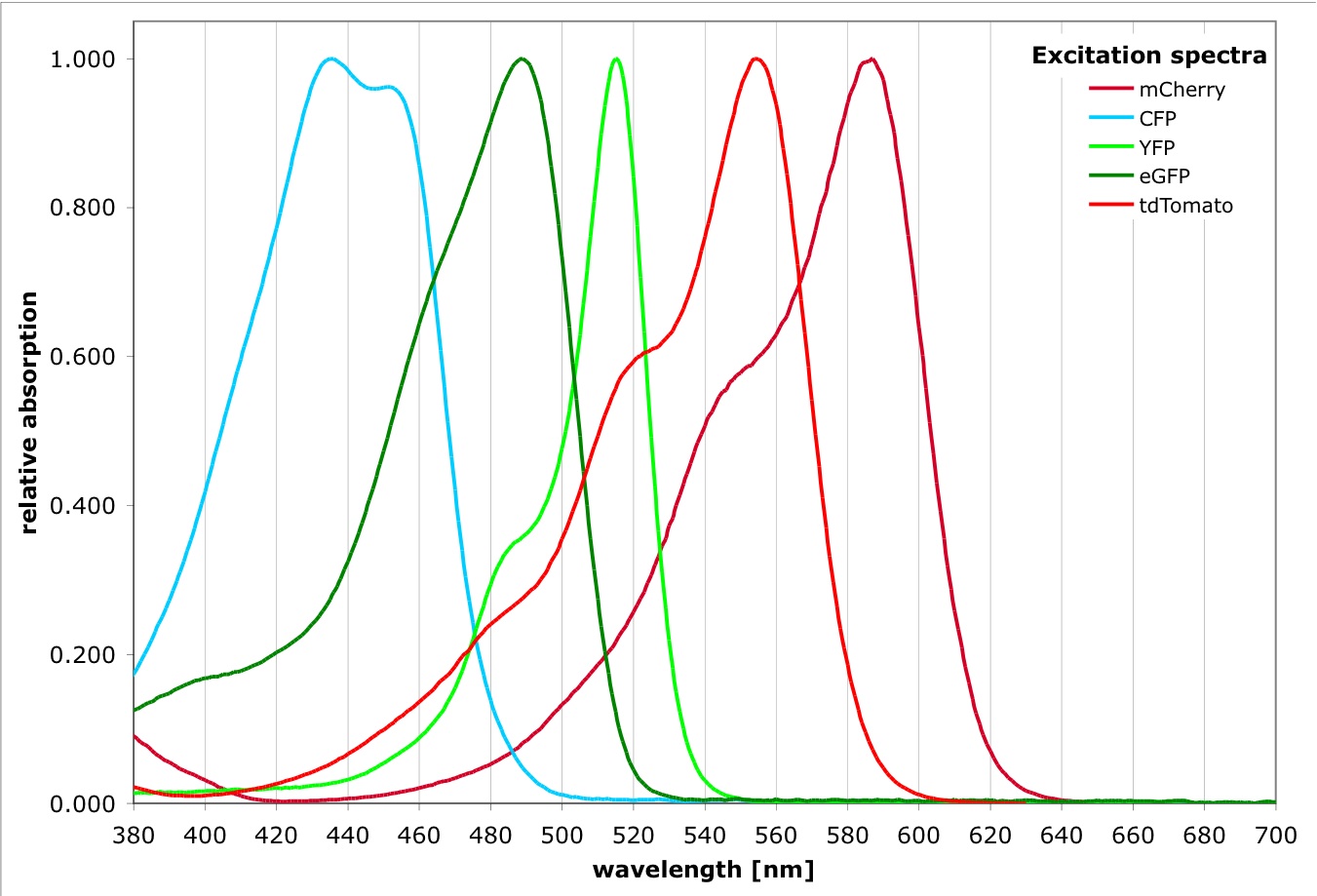
Fluorescent Protein Info:
- Excitation spectra
- Emission spectra
-
- All FPs have significant spectral overlap with other FPs!
- Make sure you use appropriate filter sets and run controls to check for cross talk.
- "Safe" combinations for co-localization are usually CFP & YFP or GFP & mCherry.
- More information on a wide variety of fluorescent proteins can be found at the Thorn lab and at the Nonet lab.
Please note that ER-g and px-g markers use mGFP5 from Jim Haseloff whereas the other green markers use sGFP (S65T).
For questions about the organelle marker set, please contact Dr. Andreas Nebenführ by email (nebenfuehr<at>utk<dot>edu).
Other Constructs
We also have created a series of fluorescent expression vectors for translational fusions. They are not included in the organelle marker set, but are described here.
|
